This is a case I treatment planned over a year ago, but due to finances the patient did not follow through after I ordered the guide. I had a pretty light day production wise and in he came wanting to get his implants. So who am I to say no?

We scanned him with a two grade duplicate denture with barium sulfate attached to the SICAT bite plate.

In the 3D view we can only see the teeth and not the flange which makes it difficult to visualize where to place the stabilization pins. You can see the flange in the cross sectional.

Go into Volumetric Mode with Contours in the 3D screen and play with the opacity of the soft tissue. Now you can see the flange. I could have scanned his denture in CEREC but I could see it well in this view.

I like my ligmaject to both anesthetize , lift the periosteum, but also to feel for flabby tissue. This guy had a very flabby ridge so I want to pay attention to whether or not my Guide moves around. Luckily his ridge was tall and there was no lateral movement.
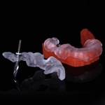
I am working with SICAT to revise how edentulous guides are made in the radiographic guide stage. IN most systems you can mount the Radiographic Guide and make the jig in purple to direct the Surgical Guide. You can’t with SICAT as the bite plate is mounted. You can make a second duplicate denture and mount that. Unfortunately I did not, but luckily there was no lateral movement of the Guide.

Looking at the occlusal of the guide it looks like the implants are too close. Thats because the Guide Sleeve has a lip that extends laterally. On the underside you can se my spacing. However he had very thin bone in the anterior Maxilla and no bone above his sinuses, so we had a narrow area Mesio Distally to get the implants in,

Nice thick ridge with a lot of tissue. We had relined his old denture prior to making the radiographic guide and in a year he did not lose much bone so everything fit very well. No rocking of the Guide.

Followed the Nobel Protocol: Punch, Counterbore, Pilot, Guided Drills:

Implants Inserted Fully Guided and Indexed:

2 #.5 by 13s, 4 4,2 by 13s, Indexed to the buccal:

Tweaked my platform positions without the Guide #4 was a little supra crestal:

Placed Impression Copings to verify Paralellism and Indexing:

Excuse the distortionin the Pan. Its hard to position a patient on a bite stick with no Anterior Teeth. I think we have a nice result for an I bar hybrid in 4 months:

