Here is a completed CEREC Guide case. I reposted the surgical pics with a little better formatting:

Patient had an existing bridge from 2-4. #2 non restorable due to decay. Sectioned bridge and fabricated a CEREC Radiographic Guide:
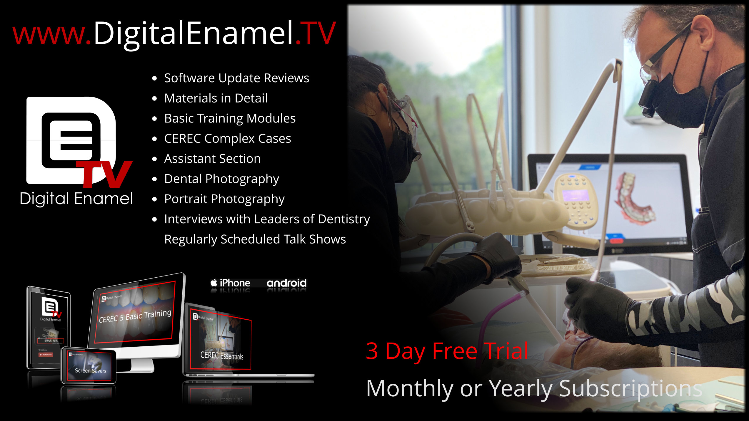 http://www.Digitalenamel.tv
Scanned the pre and the post flattened molar models in CEREC and imported without formal Proposals. I cab toggle the crowns off and on just like a conventional proposal:

Located Reference Bodies and positioned the implants. Note the position of the 7mm implant in the interspetal bone:

Milled Guides, using the Nobel Keys with the Implant Direct Drills:

You know how I roll right? Pilot through the furcation with the Guide to give you a “Glide Path” into the interseptal bone with the remaining drills:

Section that bad boy and remove each root separately with Luxators. Do everything to avoid breaking any buccal or lingual plates and preserve that interseptal bone:

Go through the series, note how well centered the osteotomy is in the socket, try doing that non-guided ;}

On to #3, same series of drills:

7mm crestal bone drill, great stability even in a socket. I have been using the HA coated lately but this one was a standard SBM surface treatment.

Standard Legacy 3 5.2 by 11.5 on #3:

In go the implants, leave the fixture mounts on, Direct Gen around the socket:

Membrane tacked down by long healing cap. Since we place these 7mm really subcrestal use a long one or you may br grinding bone off of it:

Planned and Placed:

4 months later, note the bone fill on #2:

Abutments Torqued, great solid torque values!!!

Final! Note the Implant Used as a lab analog. Usually my failed implants languish in the Box of Shame, but this time I sent it to the lab to save a few bucks. I think we got a decent result!