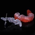
Here is a completed case that I did the implants on two separate occasions with two different guides. #14 broke, at the time my schedule was open so we did a CEREC Guide. When #15 later broke, I was swamped so I opted for an Optiguide.

Both were scanned on two different occasions with two different protocols. On #14 we located the reference body after planning with the CEREC .ssi file.

On Both cases we used 7mm implants. Drilled into the furcal area with a pilot, then sectioned and removed the roots.

After the roots were removed we proceeded as normal. On the left with the CEREC Guide and on the Right with Optiguide. Both are just as accurate. Again, these case were done months apart.

HA coated implants were used after using the crestal drill. I use HA implants on all upper molars.

Graft packed in socket and membrane tacked down with the healing abutment.

Another view on #16 tacked the membrane into the space between the gingiva and the buccal and lingual after a little reflection with a peiosteal.

This is a cool image as it shows the difference between using regular healing abutments and the contour healers. Look at how well the gingiva is reflected

Another view showing scalloping of the gingiva between the contour healers. Closed tray fixture level impression.

Stock abutments back from the lab.

Abutments seated, again look at the scalloping.

Final! Great torque values on these big 7mm. Decent profile and gingiva.
