Here is a prime example of why I do implants virtually 100% Guided. Patient came in recall and we got a stick on the distal of #3 with some sensitivity. I don’t know what gave me the forsight to take a scan, but look at the PA vs the Tangential Screen in the CBCT. Pretty obvious we are dealing with more than just a crown replacement. Got into it, the combination of a lot of decay with almost all of the clinical crown gone and really calcified canals left us with either extracting or sending the patient to the endo and perio specialists. The Patient opted for extracting and immediate implant. I was running way behind with this one, hence the lack of clinical photos.

4 months later we are ready to restore and you can see why I am not proud of this one. The angulation of the implant is definately canted and the location of the platform is way to distal. If you have been to any of my lectures you know that I hate two things, blood and mesial or distal cantilevers. No Bueno!

The abutment/impression coping of this 7mm Legacy 2 shows why I do all my cases Guided, angulation now needs to be corrected and the forces of occlusion will be at an angle.

You can see more easily now, the platform is distal and we have the dreaded mesial cantilever.

Abutment torqued. Also looking at this case the lab could have started that margin a little more apical for emergence.

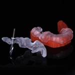
Final, my lab guy is a champ and made up for my lack of surgical awesomeness in this case. One of the things I love/hate about clinical photography is the ability to go back and improve your technique. Definitely will slow it down next time and get the CEREC Guide stuff out.
