This case is a great example of using analog and digital dentistry together to make a huge impact in a patient’s life. Duration of this case was 3.5 years as we started the process while keeping the end result in mind. Not a single drop of impression material was used on the restorative side of this case!
The patient presented with severe crowding and wanted to have nice straight teeth that he could feel confident about showing off! After consultation, a long term plan was developed and the patient agreed to go through orthodontics. After 3 years and 3 months, the orthodontics were completed and a restorative plan was made. This plan consisted of crowning 8 teeth, #’s 4-13 (5 and 12 were extracted during ortho).
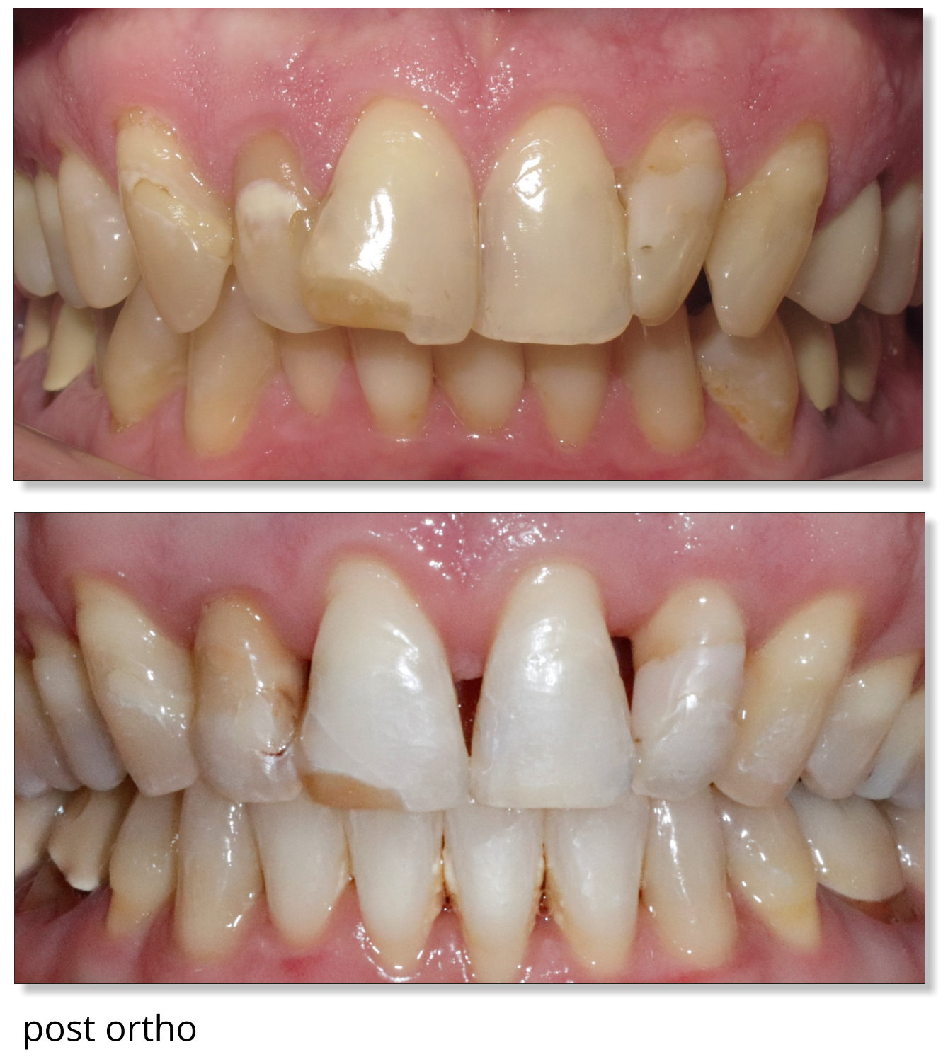
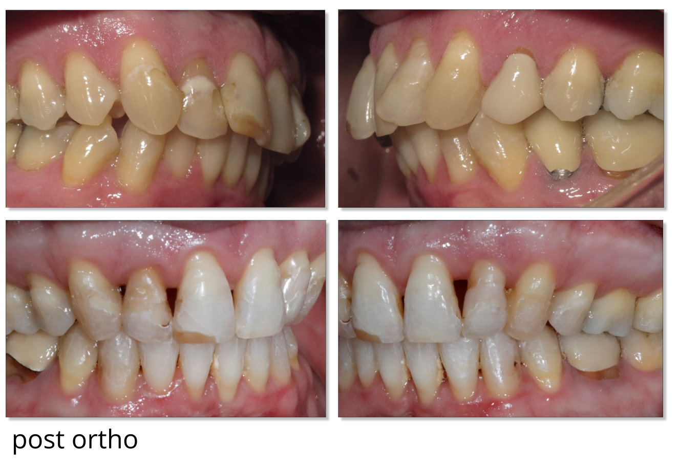
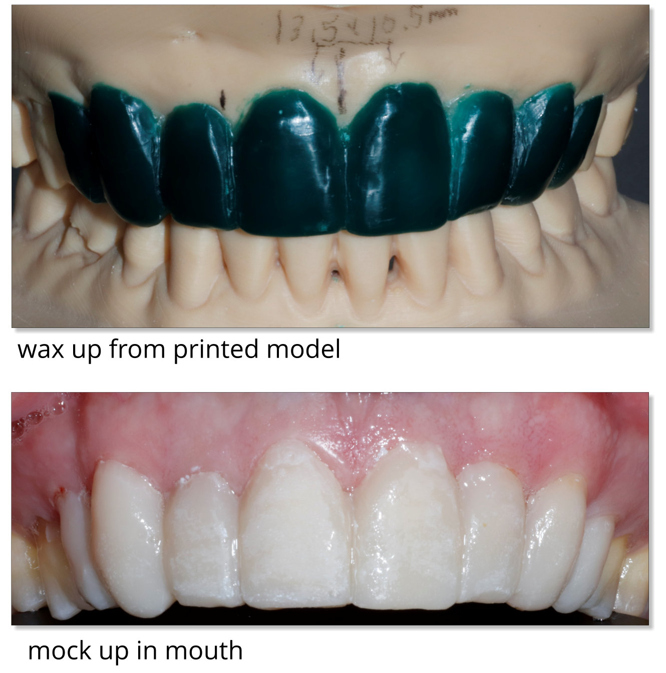
A post ortho Cerec scan was done and a .stl file was exported to print the maxilla and mandibular models. A Form 2 printer from Formlabs was utilized for model printing. A wax up was done to change the shape of the teeth. Large dark triangles opened up during the orthodontic phase so care was taken to make sure the contacts were 5mm or less to the crest of the bone to see if it was possible to gain the papilla back. After the patient approved the wax up, an intraoral mock up was done so the patient could envision the drastic change in his smile.
Preparations were done on teeth #4-13 with the mock up still on to gain all necessary clearance for the patients new smile. A bite registration was taken with a stick bite and scanned into a gingival mask catalog to relay any cant the maxilla might have to the digital models. The preparations, a bite and the opposing models were scanned into Cerec, and through Cerec connect we sent the case to Winter Springs Dental Lab, a full service digital lab. 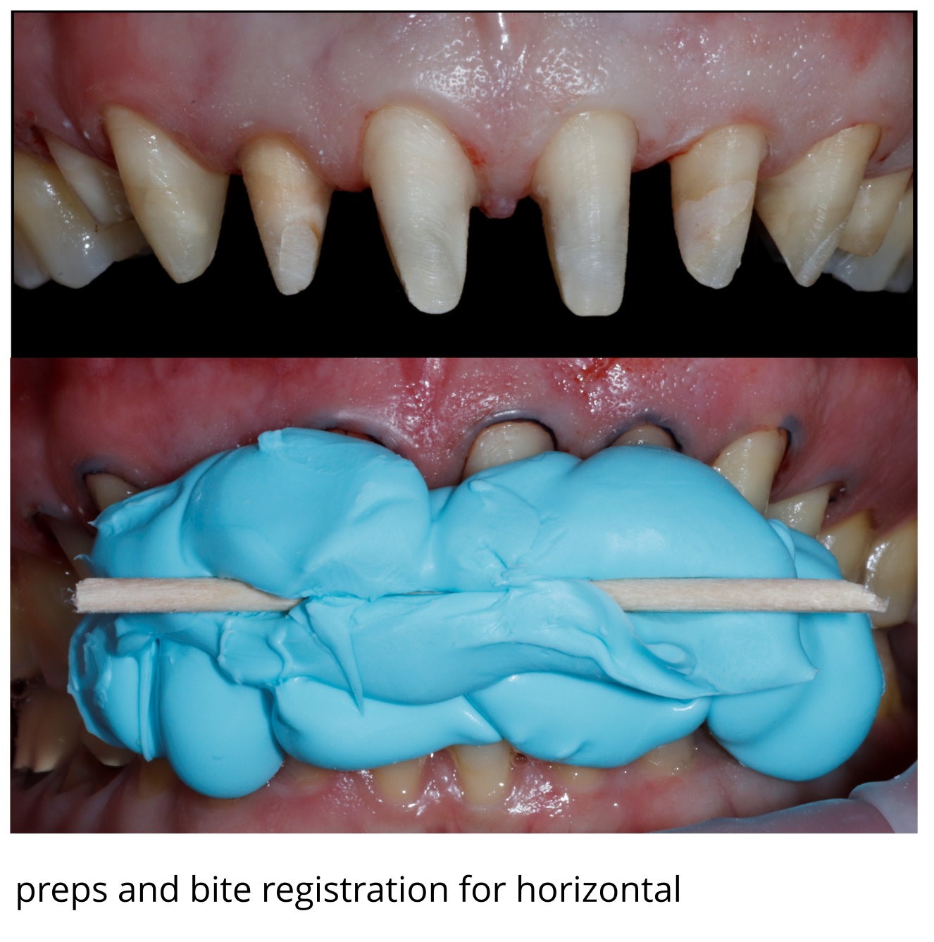 Temporaries were fabricated and used to see how the tissue would respond and to see if any changes would need to be made in contact position, length of the anterior teeth and just to make sure the patient was pleased with the look and feel of everything.
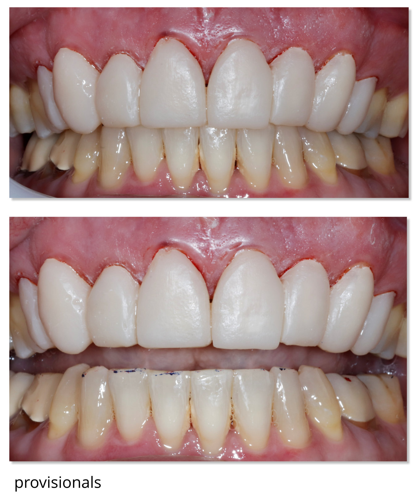
The patient returned to the try in appointment and was very pleased with the temporaries and excited to see his final restorations, which were tried in and accepted. The final restorations were cemented and photos were taken at the day of cementation to document the papilla and look of the new smile. 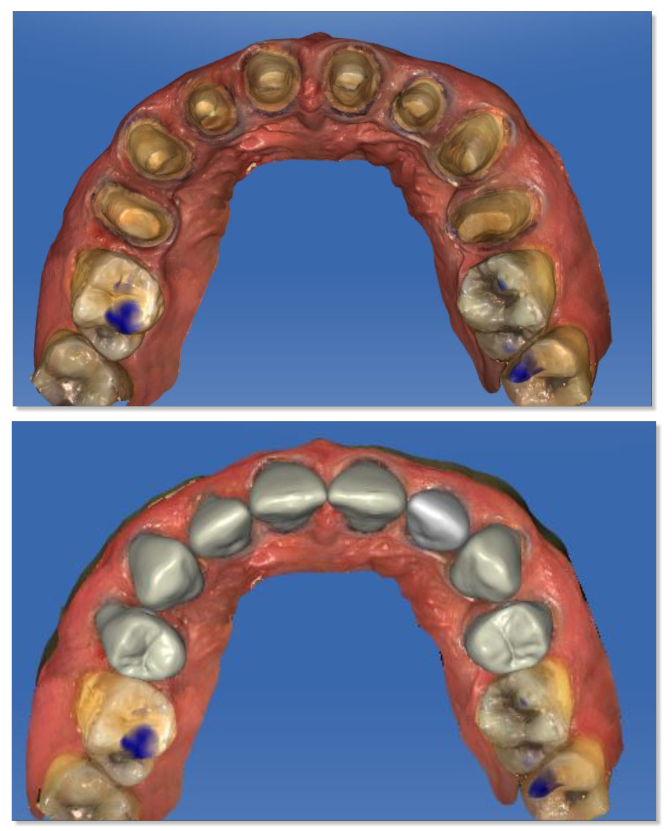
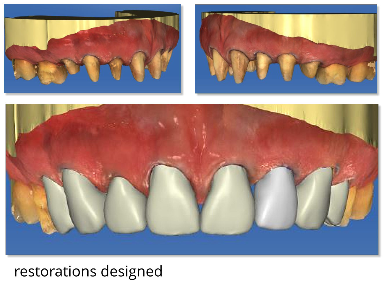
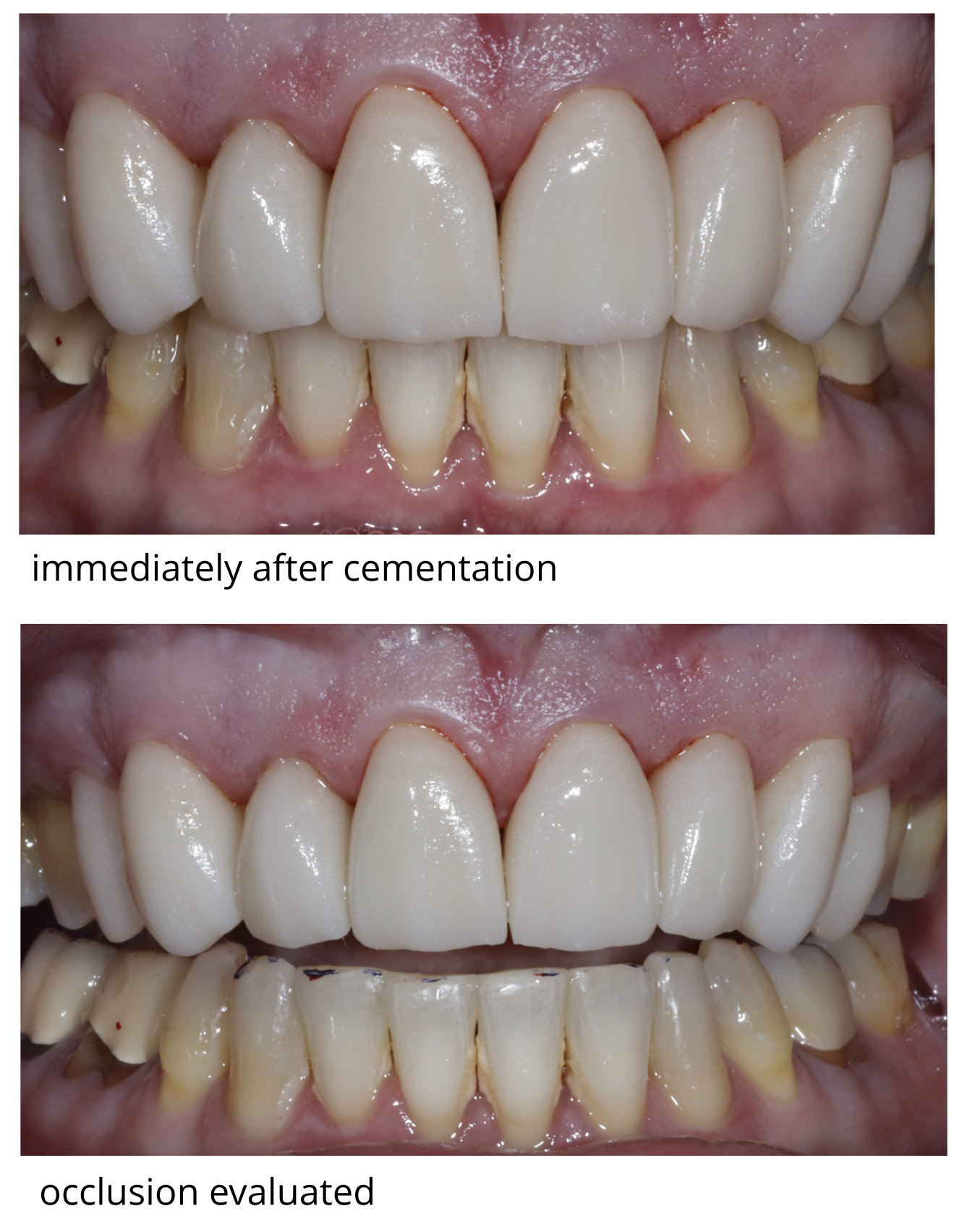
At the two week follow up appointment new photos were taken showing the papilla fill and overall gingival health. The patient’s occlusion was checked again to make any minor adjustments. No adjustments had to be made at the initial delivery appointment or follow up appointment.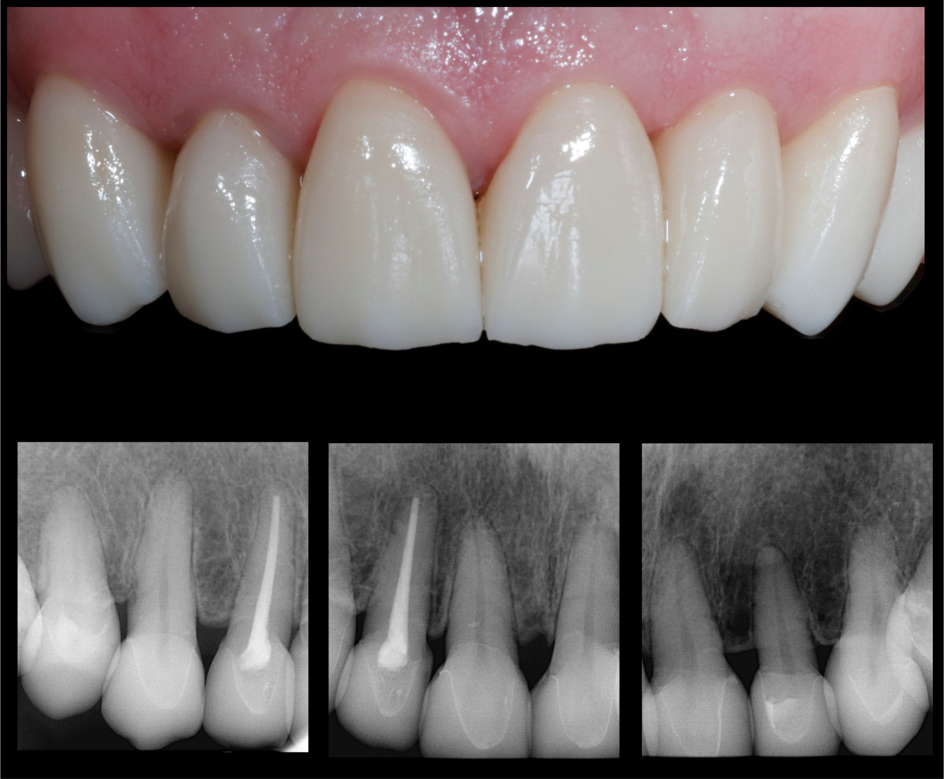 This is a great case to show that if one is not completely ready to jump into a 100% in office fabrication situation, a digital workflow can still be successfully utilized. The patient commented multiple times about not having to have a bunch of “goop” in his mouth. A proposal was done chairside with the Cerec AC unit and the restorations were close in shape and size to our lab fabricated crowns. Due to the length of the case and the papilla situation the use of Winter Springs Dental Lab was a great option, however this case could have been done successfully with chairside design and in office milling.
